Sunday, March 31, 2019

Who invented CRISPR?

There are actually two questions behind this question.
Let's start with the first question.
Who discovered CRISPR?
In 1987, Yoshizumi Ishino at Osaka University was studying the E. coli gene iap. During their sequencing efforts (remember, this was 1987), they found this odd 29 nucleotide repeat sequence with a 32 nucleotide spacing.
The final sentence of the paper [1]
So far, no sequence homologous to these have been found elsewhere in procaryote, and the biological significance of these sequences is not known.
In the post sequencing era in 2000, Francisco Mojica and Guadalupe Juez's team at the University of Alicante did a quick comparative analysis of prokaryote genomes and identified a series of Short Regularly Spaced Repeats (SRSRs) that were common among multiple species. At the time, there were speculated if it had any functional aspect or was simply an ancestral byproduct of evolution. [2]
In 2002, Ruud Jansen of Utrecht University again found a 21-37 bp repeat and citing the Ishino and Mojica work, recognized that these interspaced short sequence repeats have a distinct spacing which varies by organism. In their cited examples, Salmonella typhimurium was 21 bp where as Streptococcus pyogenes was 37 bp. Notably, they found that CRISPRs were unique to certain prokaryotes and not viruses nor eukaryotes.
Most notably, they identified a common sequence, GTT/AAC at the ends of the ends. Upstream of the CRISPR loci, there was a long homologous sequence without an open reading frame indicative of a conserved ncRNA segment. Nearby, there were able to identify homologous genes which would become cas1 to cas4. Working with Mojica, they came up with the name Clustered Regularly Interspaced Short Palindromic Repeats (CRISPR) [3]
It was then realized that Ishino happened to discover the first CRISPR loci.
Which then really made things get interesting and started several searches. What was the point of these CRISPRs?
In 2006, Eugene Koonin began hearing that several groups were finding viral DNA between the CRISPR repeats. The original hypothesis was that the CRISPR were associated with DNA repair. However, from bioinformatics searches, the huge presence of foreign DNA in spacers that were regularly getting turned over was indicative of a mechanism akin to RNAi. [4] Koonin proposed that CRISPR was a defense mechanism that enabled immunological memory.
Some microbiologists were crazy enough to listen. Rodolphe Barrangou and Philippe Horvath were working at Danisco, a yogurt company, and they were curious about strategies to help their yogurt cultures survive viral infections. They started infecting their bacteria with viruses and looked for viral DNA in the CRISPR spacers. Sure enough, the viral DNA was contained in the immune cells. When the DNA sequences were removed from the spacers, immunity was lost. In 2007, there was mechanistic proof that CRISPR served as a mechanism to defend against viruses using a completely novel mechanism of action. [5]
By 2010, the world of CRISPR molecular biology was exploding. It would be determined that the CRISPR system would behave in a certain pattern.
  • Foreign DNA would incorporated into the CRISPR loci as a spacer via CRISPR adaption.
  • The CRISPR cluster would be expressed as the pre-crRNA. The pre-crRNA would then be processed by Cas6/CasE/Csy4. The mature crRNA is bound to the Cascade complex to generate a guide RNA.
  • At that point, CRISPR interference occurs and the Cascade complex cuts the foriegn mRNA and DNA using Cas3. [6]
2005-2012 was this incredibly exciting time for CRISPR biology. It was this fascinating completely unexplored field of ncRNAs and with the right set of experiments, you could quickly open huge doors with an easy Nature paper and groups were tripping over each other to prevent getting scooped. It was one of the most exciting areas for basic research in molecular biology.
I would also say that no one was quite ready for what was going to happen next.
Which brings us to the real question
Who invented the CRISPR/CAS9 system?
Jennifer Doudna was famous not for CRISPR, but for solving one of the most challenging crystal structures, the Tetrahymena Group I ribozyme in 1991, the 2nd RNA solved structure since tRNA. Until recently, she has largely specialized on solving RNA structures. At this point, if Doudna never made CRISPR, she would have already had a respectable career probably getting into the National Academy of Science on top of a whole array of prizes that she already won before going into CRISPR research.
Because of their expertise in RNA-protein biochemistry, her group started studying RNAses like Argonaute, Dicer, and the CRISPR endonucleases. Shortly after Barrangou's discovery of the role of CRISPR, a new postdoc Blake Wiedenheft and later Martin Jinek decided that it would be neat to try to crystallize various CRISPR proteins. Very quickly, the Doudna group was able to generate structures of the multiunit CRISRP Cascade and from those structures identified the key enzymes related to cleavage.[6] In 2009, they identified Cas1 as a DNA-specific endonuclease [7] and in 2010, identified Cys4 as the Cas6 protein responsible for pre-crRNA processing. [8]
With a clear insight of the mechanism of the CRISPR/Cascade, it became clear that these Cas proteins were endonucleases that had the ability to cleave any DNA strand in a mechanism very similar to that of RNAi. Rather than relying on using all 6 Cas proteins of the Cascade, they tried identifying a smaller system.
Emmanuelle Charpentier was a new Professor who was studying trans acting genetic elements in microbes and established herself as an expert on regulatory ncRNAs. From studying Streptoccus pyogenes, her group identified an abundance of trans-activating CRISPR RNAs (tracrRNA) that would help process the CRISPR RNA using a combination of the Cas9 (at the time called the Csn1 protein) and RNase III via the type II CRISPR/Cas system. [9]
Shortly after that work in 2011, Charpentier and Doudna meet at a conference and from that meeting they decided to start a collaboration to fully understand the mechanism of the Cas9 protein. The collaboration was were strengthened by two Polish postdocs, Martin Jinek and Krzysztof Chylinski, who were excited about the opportunity to communicate with their fellow countrymen thousands of miles away. This team isolated the Cas9 protein and identified the Cas9 protein as a DNA endonuclease guided by two RNAs, the tracrRNA and the processed crRNA in a sequence specific manner contained in the crRNA. As a result, the tracrRNA not only serves a role in crRNA processing but also plays a role with the Cas9 activity itself. The double stranded RNA as well as the PAM sequence is required for Cas9 recognition of the crRNA. [10]
Through biochemical mutations in the Cas9 protein, they isolated the active sites and recognized that cleavage occurs in two domains, the HNH domain cleaves the complementary strand where as the RuvC domain cleaves the noncomplementary strand.[10]
The really key experiment was an examination of the crRNA and tracrRNA secondary structure. Recognizing the structure, a single guide RNA could be created to create a system that would be able to trigger the Cas9 endonuclease to induce double strand breaks.[10]
In that pivotal study, Doudna and Charpentier recognized through a very careful set of experiments that the Cas9 endonuclease was able to generate double strand breaks, a key finding that would allow scientists to carefully cut DNA in specific locations and trigger double strand break repair. That ability allows anyone to insert a DNA fragment of choice. [10][11] It was June 2012.
With the mechanism solved and the proof of concept demonstrated, it lead to a massive footrace to demonstrate the same in humans.
Feng Zhang was already famous because of his work in developing Optogenetics. By inserting a gene into a neuron cell, they could activate specific neuron cells simply by shining light into the position. With that ability, a scientists would be able to control the actions of a monkey purely with the switch of the light. In turn, Feng help launch an entire field of neuroscience and until 2012, was probably the breakthrough of the decade. Already at a young career, Feng was in a good spot to share a several awards with Karl Deisseroth and Edward Boyden for that body of work.
However, the huge challenge with the system was readily producing neurons with the integrated gene. As a postdoc with George Church, Feng was exploring methods on using the TALE system to rapidly and easily target DNA sequences in mammalian genomes. With the early realizations of the Cas9 endonuclease system, it was just a matter of time before a way to make it work in humans was possible. Coming from George Church's group, a group that was already well positioned to manipulate genes, Zhang was able to quickly commercialize the technique.
In January 2013, 4 sets of papers were published:
Which is where things get really complicated. Due to their experience with IP and tech transfer, Zhang was able to get the earliest and broadest patent on the CRISPR/Cas9 technology and has been able to develop the streamlined system and software that most users of the CRISPR/Cas9 technology generally use. Doudna has her own set of patents but her claims have been hampered by the existence of Zhang's patents.
Not to be outdone by each other, each group have also elucidated the crystal structures:
  • Jinek and Doudna: 6Feb14 2.6 and 2.2 angstrom structures [16]
  • Nureki and Zhang 13Feb14 2.5 angstrom structures [17]
  • Anders and Jinek: 27July14 2.6 angstrom structures. [18]
I would strongly suggest looking at this article from Quanta Magazine which seems to have done the most research on the topic. Breakthrough DNA Editor Borne of Bacteria | Quanta Magazine. HHMI's article from 2011 is also a good read When Worlds Collide.
In conclusion
CRISPR is a brand new field of biology that has multiple key discoverers.
  • Yoshizumi Ishino is credited for originally finding the CRISPR gene.
  • Ruud Jansen is the one who came up with the name.
  • Eugene Koonin is the one who proposed the purpose.
  • Rodolphe Barrangou is the one who proved the role of CRISPR.
  • Emmanuelle Charpentier is credited with identifying the importance of Cas9 and the tracrRNA.
  • Jennifer Doudna is credited with doing the biochemistry to determine the mechanism and to create the CRISPR/Cas9 system.
  • Feng Zhang is credited with the humanization and commercialization of the technique.

Wednesday, March 27, 2019

New and Upcoming Viral Vectors - Spring 2019

Since the beginning of our viral service in 2016, we’ve added many new tools to our inventory of ready-to-use viral vectors. Here are some of the AAV we have released so far in 2019. You can also browse our entire AAV inventory!
Our new AAVs include:
  • pGP-AAV-syn-FLEX-jGCaMP7b-WPRE
  • pAAV.hSyn-FLEX.iGABASnFR
  • pAAV.hSyn-FLEX.iGABASnFR.F102G
  • pAAV-Syn-Archon1-KGC-GFP-ER2
  • pAAV-Syn-FLEX-rc [Archon1-KGC-GFP-ER2]
  • pAAV-FLEX-tdTomato
  • pAAV-GFAP104-mCherry
  • pAAV-mDlx-NLS-mRuby2
  • CAG-NLS-GFP
  • pAAV-hSyn-mCherry
Read on to learn more! 


Calcium sensor AAV

The GFP-based calcium sensors are single-wavelength sensors with intensities that change in response to calcium binding. There are a few versions of these jGCaMP7 sensors with different properties (for example, overall brightness or on/off kinetics) that makes them suitable for different applications. We recently released a new variant of jGCaMP7 (keep reading) and will be releasing more variants in the next few months (see “coming soon”).
The 7b variant (pGP-AAV-syn-FLEX-jGCaMP7b-WPRE) exhibits the brightest resting fluorescence of the GCaMP7 family of calcium sensors and can be used for imaging of small neuronal processes like dendrites and axons.
See all calcium sensor AAV at Addgene.

GABA sensor AAV

GABA is the primary inhibitory neurotransmitter in vertebrates, but methods for directly imaging GABA have been limited. Like calcium sensors, different iGABASnFR have been developed and are suited for different applications. The iGABASnFR GABA sensor (pAAV.hSyn-FLEX.iGABASnFR) has good membrane localization and brightness. The F102G variant (pAAV.hSyn-FLEX.iGABASnFR.F102G) is noticeably dimmer than iGABASnFR, and accumulates in the endoplasmic reticulum, but exhibits larger fluorescent responses to GABA.
Find the iGABASnFR and its F102G variant in AAV1 at Addgene!

Archon1, a voltage sensor AAV

Historically, electrical activity in the brain was measured with implanted electrodes. The Archon1 voltage reporter allows a way to monitor neuronal electrical activity through imaging. Since it’s illuminated by red/orange light, Archon1 can also be used in conjunction with optogenetic activators and inhibitors that respond to blue light. We recently released a constitutive (pAAV-Syn-Archon1-KGC-GFP-ER2) and cre-dependent version (pAAV-Syn-FLEX-rc [Archon1-KGC-GFP-ER2]) of this membrane-embedding voltage detector.
Find Archon1 and its cre-dependent version at Addgene!

Fluorescent protein and control AAV

These fluorescent protein expression vectors can be used to visualize cells and to quantify targeting of particular cell types. For example, the mDlx enhancer restricts expression to GABAergic interneurons. These vectors can also be used as the control vectors for experiments encoding a fluorescent-tagged molecular tool.  

Viral vectors coming soon!

These vectors listed below should be released at Addgene in the next 6 months, pending quality control. For an estimate on availability date or to be notified when a particular catalog item is available for order please email help@addgene.org with the vector name and serotype you’d like to be notified about.

Controls

PlasmidSerotypeName
114472 AAV5, AAV8 pAAV-hSyn-mCherry
50459 AAVrg pAAV-hSyn-DIO-mCherry
59462 AAV2 pAAV-CAG-tdTomato (codon diversified)
37825 AAV8*, AAV9* pAAV-CAG-GFP
50465 AAV8*, AAV9* pAAV-hSyn-EGFP
*Also coming in our new 20 ul trial size!

Voltage indicators

PlasmidSerotypeName
119036 AAV1 pAAV-hsyn-flex-Voltron-ST

Chemogenetics

PlasmidSerotypeName
75033 AAV1 pAAV CD68-hM4D(Gi)-mCherry (CD68 is a 
 microglia/macrophage promoter)
121539 AAV5 pOTTC1596 - pAAV SYN1 HA-hM3D(Gq)
121538 AAV5 pOTTC1484 - pAAV SYN1 HA-hM4D(Gi)
119741 AAV5 AAV SYN flex PSAM4 GlyR IRES EGFP
 Chimeric channels for neuronal manipulation
119742 AAV5 AAV SYN PSAM4 GlyR IRES EGFP
 Chimeric channels for neuronal manipulation

Recombinases

PlasmidSerotypeName
50363 AAV5 AAV phSyn1(S)-DreO-bGHpA
121675 AAV9 pAAV-EF1a-fDIO-Cre (FLP-dependent CRE)
55634 AAV1 pAAV-EF1a-mCherry-IRES-Flpo
55637 AAV1 pAAV-EF1a-Flpo

Calcium sensors

PlasmidSerotypeName
104496 AAV1
 GP-AAV-CAG-FLEX-jGCaMP7f-WPRE AAV1 with a CAG
 promoter exhibits high specificity for Purkinje neurons.
105321 AAV1
 pGP-AAV-syn-jGCaMP7c variant 1513-WPRE 7c exhibits high
 contrast between peak fluorescence and resting fluorescence.
 It is useful for activity imaging of large populations of densely
 labeled neurons because background fluorescence from
 inactive neurons is reduced.
105322 AAV1
 pGP-AAV-syn-FLEX-jGCaMP7c variant 1513-WPRE7c exhibits
 high contrast between peak fluorescence and resting
 fluorescence.  It is useful for activity imaging of large
 populations of densely-labeled neurons because background
 fluorescence from inactive neurons is reduced.


Friday, March 22, 2019

Tips for a 1st time AAV user (by a Rookie AAV user)

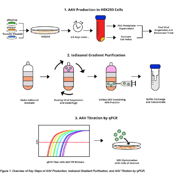
My lab's vector of choice is AAV, with nearly every experiment requiring AAV. Before joining my lab, I had never worked with AAV, so naturally I had to package some virus for my first experiment. It was a bit intimidating, but I had my lab’s protocols and some great co-workers to help me out. Even with these tools, I found myself writing AAV production tricks into the margins of my protocol. While these tips weren’t critical to the experiment, they definitely made my life easier!  In this post, I’ll share some AAV productionpurification, and titration tips, while also summarizing the basic steps and analyses needed for packaging AAV.
AVV production, purification, and titration


AAV production

Overview: The first step in packaging AAV is transfecting HEK293 cells with AAV packaging plasmids. The cells need three different plasmids to produce AAV: 1) the RepCap plasmid which provides the AAV replication (rep) and capsid (cap) genes. AAV replication uses the host’s polymerase, but requires Rep proteins to process a double-stranded intermediate into the single-stranded genome that's then packaged into AAV’s protein shell, or capsid; 2) the pHelper plasmid which expresses adenovirus genes which help mediate AAV viral replication; and 3) the transfer plasmid which encodes a transgene of interest that’s packaged into the virus. Two to five days after transfection, the AAV-containing cells and media are harvested and purified. In total, this process takes 4-7 days, not counting the time needed to expand the HEK293 cells. Check out the Addgene AAV Production protocol for more details.
Pro tips
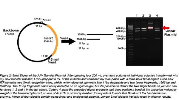
  1. I’m not going to lie, AAV transfer plasmids are kind of a pain to work with. These plasmids contain ITRs which are required for proper packaging of AAV, but also form secondary structures that are prone to deletion from the transfer plasmid. The good news is there’s a few simple ways to deal with this issue. One way is to grow multiple bacterial cultures of your AAV transfer plasmid at 30 °C instead of 37 °C and then screen for ITR recombination with a SmaI or XmaI restriction enzyme digest (the ITRs contain SmaI and XmaI restriction sites). Another way is to transform AAV transfer plasmids into bacterial strains, like NEB Stable. NEB Stable competent cells lack RecA, a protein that aids in the homologous recombination of duplicated regions like the ITRs. Restriction digest screening for ITR deletion is still necessary.

    AAV plasmid SmaI digest

  2. To save time and money, I like to miniprep 5 mL of my AAV transfer plasmid cultures and then screen with SmaI digests. I freeze the bacterial pellet for the remainder of the cultures and later maxiprep only the cultures with intact ITRs.

  3. I use a spreadsheet similar to this one to plan my AAV packaging PEI transfections. It saves me time and helps me figure out if I have enough plasmid to complete my experiment.

  4. AAV is usually the limiting reagent for my experiments, so I always make more AAV than I think I need. Unlike lentivirus, AAV requires a higher number of viral particles for efficient gene transfer. I figure it’s better to have a bit extra AAV than to have to produce more virus because I don’t have enough AAV.

  5. Compared to the Addgene AAV Production protocol, I take a lazier approach for harvesting my AAV. I skip the PEG precipitation of the HEK293 cell culture supernatant because it’s time consuming to prepare the 40% PEG solution. PEG takes several hours on a heated stir plate to dissolve and requires monitoring so that the solution doesn’t get too hot because then the PEG will separate into two phases. If this happens, the solution can be cooled and the two phases mixed together. Lastly, the PEG solution needs to be sterilized, either by filtration, which takes time because the solution is viscous, or by autoclaving, which also takes time. Rather than investing all of this effort in preparing reagents, I choose to only harvest the cell pellet. My yield would be higher if I used PEG precipitation, but for my experiments, I get enough virus from the cell pellet alone. If yield is important, or if the viral particles of the AAV serotype you’re working with are primarily found in the culture media, you should consider performing PEG precipitation.

  6. Instead of sonicating the cell pellet, I lyse the cells with this AAV lysis buffer and freeze/thaw the lystate four times in a 95% ethanol and dry ice bath to release the virus particles from the cells. Cell lysates are then treated with benzonase immediately before iodixanol purification. This approach gives me a bit of flexibility since I can store the cell lysates at -80 °C for up to 6 months before purifying the AAV (Choi et al).

AAV purification with iodixanol gradient ultrafugation
Overview: Iodixanol gradient ultracentrifugation uses a gradient of different concentrations of iodixanol to separate out contaminants from an impure AAV preparation. 15%, 25%, 40%, and 60% iodixanol solutions are carefully layered and then the viral suspension generated during AAV production is overlaid. Following ultracentrifugation, the AAV-containing 40% fraction is collected and buffer exchanged to remove the iodixanol and concentrate the purified virus. This process can be completed in one long day, or the virus can be stored at 4 °C and buffer exchanged the next day. Refer to the AAV Iodixanol Gradient Ultracentrifugation Protocol for more details.
Pro tips
  1. Watch this AAV purification video! It didn’t exist when I did my first iodixanol gradient purification, but I wish it had. The video gives a brief overview of how iodixanol purifications works, shows how to create the iodixanol gradient layers, and has some great pointers that while help you master iodixanol AAV purification.

  2. If you’re new to creating gradients, practice making the iodixanol layers before doing your first purification. This helped me get a feel for layering the iodixanol solutions and let me double-check that the iodixanol gradient solutions were prepared correctly.

  3. When aliquoting purified and buffer exchanged virus, remember to make one or two small aliquots to use for qPCR titering. This helps avoid multiple freeze/thaws of your larger aliquots of virus.

AAV titration by qPCR

Overview: AAV titering by qPCR quantifies the number of genome-containing viral particles that are present in an AAV prep. AAV samples are first DNaseI digested to remove any residual AAV plasmid that was carried over from the AAV production process. Either SYBR green technology or a TaqMan primer/probe set can be used and samples are quantified by comparing to an AAV standard curve, which is generated from an ITR-containing plasmid. This whole process takes ~3 hours to complete: 1 hours hands-on time and 2 hours for the qPCR run and data analysis. Refer to this Addgene AAV Titration by qPCR Protocol for more details.
Pro Tips
  1. When calculating the titer for your AAV, remember to account for the dilution of the sample for the DNAseI digest.

  2. Use good sterile technique when handling your AAV plasmid standard and AAV transfer plasmids. It’s easy to contaminate a workspace with plasmid, not that I would know about this… A no template control (NTC) should always be included.

  3. While the physical titer generated by qPCR is useful, I test a few different concentrations of virus, or multiplicity of infections (MOIs), to determine the optimal dose of AAV for my cells of interest. Every batch of AAV requires its own MOI optimization to account for batch-to-batch variability. Check out this post to learn more about different ways to titer AAV.
Do you have any AAV production tips or tricks? Please share them in the comments below!

Wednesday, March 20, 2019

Stabilised Bacterial Promoters: Constant Gene Expression at any Copy Number

Researchers express genes of interest from plasmids in order to study gene function or to engineer cells for specific purposes. Unfortunately, plasmid copy numbers vary within cell populations and over time resulting in variable gene expression that can impact observed phenotypes. Factors such as the growth medium, growth temperature, and growth rate can all impact plasmid copy number in a cell.
One can attempt to eliminate fluctuations due to copy number by introducing genes of interest into the chromosome. But this has its own set of problems. Rapidly dividing cells initiate genome replication more than once per cell division and thus, genes closer to the origin of replication will be over-represented compared to those further away.
As a result, variable gene expression can make data hard to interpret or can interfere with the precision needed in metabolic engineering or other synthetic biology circuits. What is a molecular biologist to do? Let’s take a look at how plasmids recently deposited by Chris Voigt’s lab help keep gene expression stable regardless of copy number (Segall-Shapiro et al., 2018).

Designing stabilized promoters

Expression_regulation_Figure1Badapted-01
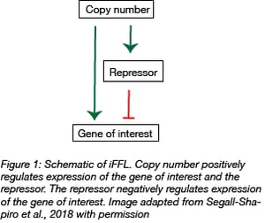
To keep gene expression stable regardless of copy number, the team made use of stabilized promoters. In contrast to unregulated promoters that are always on, stabilized promoters incorporate feedback loops such as the incoherent feedforward loop (iFFL). In general, iFFLs use an input signal that both positively and negatively controls the output. In this case, plasmid copy number is the input and gene expression is the output.

The input, plasmid copy number, directly influences gene expression. By itself, increasing the copy number of a gene increases its expression. However, if you introduce negative regulation by using a promoter that is responsive to a repressor protein, the increase in expression due to copy number is kept in check. In such an iFFL, as plasmid copy number increases, the amount of repressor protein also increases thereby intensifying repression of the gene of interest and keeping its expression level constant.
Figure_2A-01-1
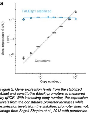
In the case of the stabilized promoters from Segall-Shapiro et al., they chose a TALE protein as the repressor because TALEs can be programmed to bind tightly to arbitrary DNA sequences and have been shown to achieve ~100 fold repression in Ecoli. In addition, TALEs bind non-cooperatively to the promoter region, which is predicted to be required to decouple gene expression and plasmid number.

In a suite of plasmids modified from pSC101 with copy numbers ranging from 3-100 per cell, a stabilized promoter buffered against the effects of copy number on reporter gene expression. On the other hand, using a constitutive promoter in the same background resulted in a 20-fold variation of gene expression.
Because pSC101 is not representative of all types of plasmids, the stabilized promoters were also tested in different plasmid backbones that varied in size or carried different genes. In these cases, gene expression also remained stable regardless of plasmid copy number.

Stable gene expression whether growing slowly or quickly

Growth media and growth stage can both affect gene copy number. Moreover, gene copy number is affected differently by growth rates when a gene is expressed from a plasmid as opposed to the chromosome. Let’s take a look at how the stabilized promoters perform during these types of fluctuations.
When cells are dividing rapidly, multiple rounds of replication are initiated before the cell has divided. This means that genes located near the origin will be over-represented compared to those further away. While constitutive promoters drove maximal gene expression near genomic origins of replication, the stabilized promoters created by Segall-Shapiro et al. showed nearly no position-dependent differences in gene expression.
On the other hand, slow growth can also cause changes in plasmid copy number. During the shift from exponential to stationary phase, the number of plasmids per cell increases 4-5 fold (for pUC plasmids) or ~2 fold (for p15a, R6K, and ColE2 plasmids) as cell division slows (Kittleson et al., 2011). Segall-Shapiro et al. showed that the stabilized promoters were able to better modulate gene expression during shifts in growth rate as compared to a constitutive promoter.
To simulate the increase in plasmid copy number during the transition to stationary phase, Segall-Shapiro et al. placed a trans-acting RepA protein under the control of IPTG. Because RepA is a plasmid replication initiator protein, they could alter copy number of the plasmid by altering IPTG concentrations. Higher IPTG concentration leads to higher RepA expression which results in high plasmid copy number. When they increased concentrations of IPTG, a constitutive promoter upstream of GFP increased expression of GFP six-fold, but a stabilized promoter increased expression only two fold. Moreover, gene expression levels returned to the baseline after 3 hours with the stabilized promoter while gene expression from the constitutive promoter did not.

Similar levels of expression between a plasmid and the chromosome

The team also showed that the levels of gene expression from stabilized promoters were equivalent whether the genes were expressed from the genome or a plasmid. Thus, a stabilized promoter can allow a system to be moved from a plasmid to the genome without greatly impacting the level of gene expression.
For example, the synthesis of deoxychromoviridans requires three genes whose proteins are needed sequentially. When Segall-Shapiro et al. placed the genes under the control of a constitutive promoter and then moved them into the genome, deoxychromoviridans production titers decreased. But when the genes were placed under the control of a stabilized promoter, deoxychromoviridans titers were similar regardless of where the genes were expressed.

What’s the use of stable gene expression?

Constant gene expression can enable more confidence in gene expression readouts from assays such as qPCR or microarray. But, aside from facilitating the joys of a p-value < 0.05, having such fine control of gene expression is useful for many metabolic engineering and synthetic biology goals. In metabolic engineering, the expression of enzymes in a pathway will ideally be optimized to promote generation of the desired product while avoiding the accumulation of toxic intermediates or the shuttling of the intermediates into other pathways. In cases where a specific ratio of products is needed, such as within a multi-protein complex, stabilized promoters can eliminate variability between the different components. When your pathway needs precise fine-tuning of gene expression levels, give these stabilized promoters a try.